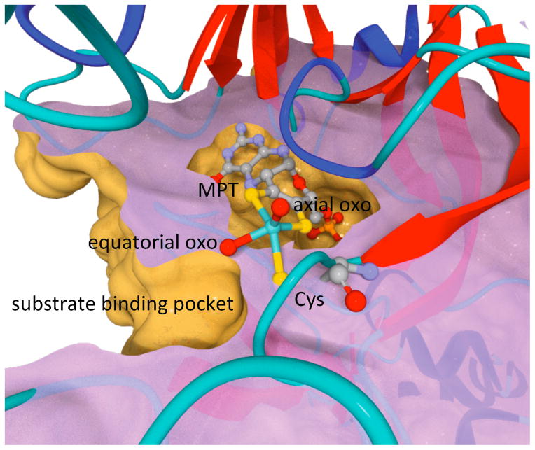
Figure 1.
Active site of SO rendered from the 1.9 Å chicken SO structure, pdb 1SOX.6 The molybdopterin (MPT), the conserved Mo-bound Cys residue (C185 and C207 in chicken and human SO, respectively), the axial and equatorial oxo ligands, and Mo are shown as ball-and-stick (blue-green = Mo, red = O, yellow = S, gray = C, orange = P, and purple = N). The protein is displayed as ribbons and a cross-section to reveal the surfaces of the substrate and cofactor pockets and their relative orientations.

