
Fig. 5.
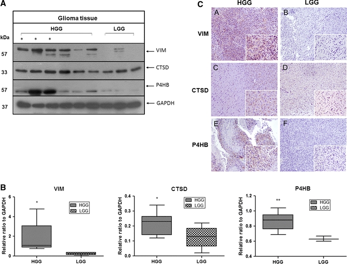
Clinical significance of VIM, CTSD and P4HB in human malignant glioma. a Representative blot images demonstrate high expression levels of VIM, CTSD and P4HB in high-grade (HGG) compared with low-grade gliomas (LGG). Glioblastoma reveals the highest level of expression of the three proteins and is illustrated by asterisk. b Whiskers (min to max) diagram shows the densitometry data from Western blot analysis. Differences are expressed as ratio of the target protein in different grade of glioma to glyceraldehyde-3-phosphate dehydrogenase (GAPDH) density. c VIM, CTSD and P4HB detection by IHC. Representative immunohistochemical staining of VIM, CTSD and P4HB in paraffin-embedded human glioma tissues. Low-grade glioma tissues seen in b, d, f are weakly to moderately stained, while in high-grade tissues, VIM, CTSD and P4HB stainings are strongly intense (A, C, E). Original magnification: ×100; ×400 (insets)