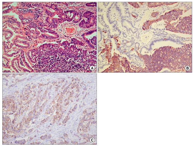
Fig. 1.
Histological finding of H&E stain and immunohistochemistry. (A) Adenocarcinoma with neuroendocrine features (H&E stain, ×200). (B) Positive in area of neuroendocrine tumor (Gold color) (Synaptophysin stain, ×200). (C) Positive in area of neuroendocrine tumor (Gold color) (Chromogranin A stain, ×200).

