Abstract
A debate is currently ongoing about whether a large gastrointestinal stromal tumor (GIST) should be treated by the laparoscopic approach because of the increased risk of tumor rupture during manipulation of the tumor with laparoscopic instruments and the resultant peritoneal tumor dissemination. Herein, we report a case of a large GIST of the stomach which was successfully treated by the laparoscopic approach. A 57 year old female patient visited our institution complaining of postprandial epigastric discomfort. An esophagogastroduodenoscopy and an abdominal computed tomography scan revealed a 10×8 cm sized submucosal tumor at the greater curvature side of the gastric antrum. The patient underwent laparoscopic distal gastrectomy with intracorporeal Billroth-II reconstruction without any breakage of the tumor. Her postoperative course was uneventful and she was discharged on the 7th postoperative day. Even a large GIST of the stomach can safely be treated by the laparoscopic approach when it is performed with proper techniques by an experienced surgeon.
Keywords: Gastrointestinal stromal tumor, Stomach, Laparoscopy
Introduction
Gastrointestinal stromal tumor (GIST) is the most common mesenchymal tumor of the gastrointestinal tract and it is most frequently found in the stomach. Fifty to sixty percent of GISTs are found as a localized disease at presentation, and 95% can be completely resected. Surgical resection with a 1~2 cm negative resection margin is the standard treatment for a non-metastatic GIST. The most important oncologic principle while resectioning is to avoid rupturing the tumor during manipulation in order to circumvent peritoneal dissemination of the tumor which can result in poor prognosis of the patient, because the characteristic of the mass is soft and fragile. The tumor commonly metastasizes to the liver or the peritoneum but it rarely metastasizes to the lymph nodes. Therefore, lymphadnectomy is not needed during surgery. (1-4) While minimally invasive surgery has recently been gaining popularity in the resection of GISTs, it lacks evidence-based recommendation, especially in the resection of a GIST larger than 5 cm.
Herein, we report a case of a large GIST of the stomach which was successfully treated by the laparoscopic approach in order to investigate the technical feasibility and the oncologic safety of laparoscopic resection for large GISTs.
Case Report
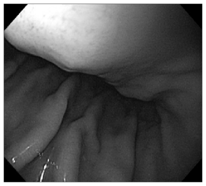
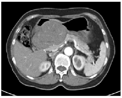
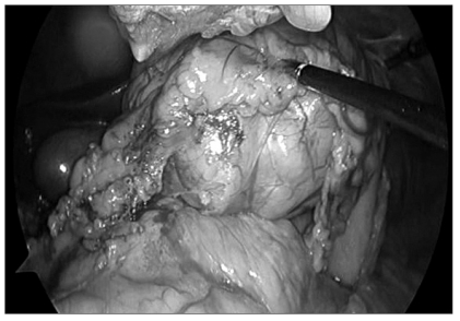
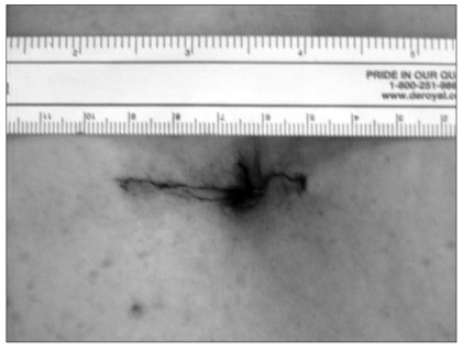
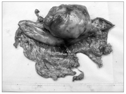
A 57 year old female patient visited the Department of Surgery, Incheon St. Mary's Hospital complaining of postprandial epigastric discomfort. An esophagogastroduodenoscopy (EGD) and abdominal computed tomography (CT) scan were performed. On EGD finding, a submucosal tumor was found at the posterior wall of the gastric antrum (Fig. 1), and it was revealed by CT scan to be a 10 ×8 cm sized heterogeneously enhanced, well circumscribed solid mass arising from the posterior wall of the gastric antrum (Fig. 2). There was no metastatic lesion in other organs. She was recommended an open resection, but insisted on laparoscopic resection. Therefore, she underwent laparoscopic resection after providing informed consent. Under general anesthesia, the patient was placed in the reverse Trendelenburg position with her legs apart. The operator was positioned on the right side, and the first assistant was positioned on the left side of the patient. The camera operator was positioned between the legs of the patient. A 10 mm trocar was inserted through the umbilicus for the camera. A 5 mm trocar and a 12 mm trocar on the right upper abdomen were used as working channels for the operator. Another 5 mm trocar and a 12 mm trocar on left upper abdomen were used for the first assistant. The greater omentum was opened with ultrasonic shears and the mass was identified. Great care was taken to avoid touching the mass directly with the laparoscopic instruments. There was no direct invasion to the pancreas (Fig. 3). The right gastroepiploic vessels were identified and doubly ligated with clips and the duodenum was resected with a laparoscopic linear stapler. The right gastric vessels and descending branches of the left gastric artery were identified and doubly ligated. The stomach was transected at 4 cm proximal from the upper border of the mass with a laparoscopic linear stapler. The specimen was placed into a plastic bag and retrieved through the umbilical port site which was extended to a 4 cm length (Fig. 4). As the consistency of the mass was soft, even a 10 cm sized mass could be extracted through a 4 cm length incision without any breakage (Fig. 5). The extended umbilical port site was partially repaired and pneumoperitoneum was reestablished. Intracorporeal Billroth-II reconstruction was performed. Oral feeding was started from the second postoperative day and the patient was discharged on the 7th postoperative day without any complication. The pathologic result was CD 117 and CD 34 positive gastrointestinal stromal tumor with 1 mitosis per 50 high power field (intermediate risk).
Fig. 1.
An esophagogastroduodenoscopy finding of gastric gastrointestinal stromal tumor.
Fig. 2.
Abdominal computed tomography findings of gastric gastrointestinal stromal tumor. A 10×8 cm sized homogeneously enhanced, well circumscribed mass which developed from the posterior wall of the gastric antrum was observed.
Fig. 3.
An operative view of gastric gastrointestinal stromal tumor. After entering the lesser sac, a 10 cm sized huge mass was noted. There was no pancreatic invasion.
Fig. 4.
Surgical wound. After resection, the specimen was placed into a plastic bag and retrieved through the umbilical port site which was extended to 4 cm length.
Fig. 5.
Gross finding. Even though, the size of the mass was 10 cm, the mass was successfully resected and retrieved without any breakage.
Discussion
Gastric wedge resection with a proper safety margin is the preferred type of surgery in treating gastric GIST. However, the type of surgery can differ according to the size and location of the tumor. In some cases in which the tumor is located at the prepyloric area or around the esophagogastric junction, more extended surgery such as a partial gastrectomy or even a total gastrectomy can be necessary.(4-6) As the laparoscopic approach is gaining popularity in the treatment of gastric GIST, many debates on its oncologic safety and relevance have arisen. The reported early postoperative outcomes of laparoscpic approaches have been comparable to or even better than those of the conventional open approach.(7) Several reports have been presented about the comparable longterm oncologic outcomes of the laparoscopic approach for GIST. However, most of these reports focus on the oncologic outcomes of small or medium sized (less than 5 cm) GISTs.(8) According to the National cancer care network guideline which was revised in 2010, Gastric GISTs 5 cm or smaller may be removed through laparoscopic wedge resection and the tumor should be removed in a protective plastic bag. However, in the case of a GIST of more than 5 cm in size, the laparoscopic or hand-assisted approach was recommended because of the benefit of tactile feedback.(9) The reason for this recommendation is that if the size of the tumor is larger, the risk of rupture of the pseudocapsule by laparoscopic manipulation would be greater.(2)
Recently, an increasing number of reports have been presented for safe laparoscopic resection for larger GISTs due to the advancement of the laparoscpic technique. However, many reports still recommend open conventional resection for tumors which are more than 7~10 cm in size.(2,5) In our case, the authors successfully resected a large gastric GIST of 10 cm in size without any breakage of the tumor using a meticulous laparoscopic surgical technique.
To avoid tumor rupture, an attached fibrosis band was left on the tumor when resecting around the tumor, and if traction was needed, the fibrous tissue or normal gastric wall around the mass was used. As a result, there was no direct manipulation of the tumor during the operation. Moreover, the size of the minilaparotomy incision could be minimized to 4 cm in extracting the 10 cm sized mass. This was possible because the characteristic of the GIST mass was soft and fragile.
The laparoscopic approach is feasible for a large GIST of the stomach when proper laparoscopic techniques are used by an experienced surgeon. However, further study will be needed on the limitation of the size of the tumor in laparoscopic resection of GIST.
References
- 1.Basu S, Balaji S, Bennett DH, Davies N. Gastrointestinal stromal tumors (GIST) and laparoscopic resection. Surg Endosc. 2007;21:1685–1689. doi: 10.1007/s00464-007-9445-z. [DOI] [PubMed] [Google Scholar]
- 2.Gervaz P, Huber O, Morel P. Surgical management of gastrointestinal stromal tumours. Br J Surg. 2009;96:567–578. doi: 10.1002/bjs.6601. [DOI] [PubMed] [Google Scholar]
- 3.Melstrom LG, Phillips JD, Bentrem DJ, Wayne JD. Laparoscopic versus open resection of gastric gastrointestinal stromal tumors. Am J Clin Oncol. 2011 doi: 10.1097/COC.0b013e31821954a7. [Epub ahead of print] [DOI] [PubMed] [Google Scholar]
- 4.Ke CW, Cai JL, Chen DL, Zheng CZ. Extraluminal laparoscopic wedge resection of gastric submucosal tumors: a retrospective review of 84 cases. Surg Endosc. 2010;24:1962–1968. doi: 10.1007/s00464-010-0888-2. [DOI] [PubMed] [Google Scholar]
- 5.Goh BK, Chow PK, Chok AY, Chan WH, Chung YF, Ong HS, et al. Impact of the introduction of laparoscopic wedge resection as a surgical option for suspected small/medium-sized gastrointestinal stromal tumors of the stomach on perioperative and oncologic outcomes. World J Surg. 2010;34:1847–1852. doi: 10.1007/s00268-010-0590-5. [DOI] [PubMed] [Google Scholar]
- 6.Catena F, Di Battista M, Fusaroli P, Ansaloni L, Di Scioscio V, Santini D, et al. Laparoscopic treatment of gastric GIST: report of 21 cases and literature's review. J Gastrointest Surg. 2008;12:561–568. doi: 10.1007/s11605-007-0416-4. [DOI] [PubMed] [Google Scholar]
- 7.Novitsky YW, Kercher KW, Sing RF, Heniford BT. Long-term outcomes of laparoscopic resection of gastric gastrointestinal stromal tumors. Ann Surg. 2006;243:738–745. doi: 10.1097/01.sla.0000219739.11758.27. [DOI] [PMC free article] [PubMed] [Google Scholar]
- 8.Ishikawa K, Inomata M, Etoh T, Shiromizu A, Shiraishi N, Arita T, et al. Long-term outcome of laparoscopic wedge resection for gastric submucosal tumor compared with open wedge resection. Surg Laparosc Endosc Percutan Tech. 2006;16:82–85. doi: 10.1097/00129689-200604000-00005. [DOI] [PubMed] [Google Scholar]
- 9.Demetri GD, Benjamin RS, Blanke CD, Blay JY, Casali P, Choi H, et al. NCCN Task Force. NCCN Task Force report: management of patients with gastrointestinal stromal tumor (GIST)--update of the NCCN clinical practice guidelines. J Natl Compr Canc Netw. 2007;5(Suppl 2):S1–S29. [PubMed] [Google Scholar]


