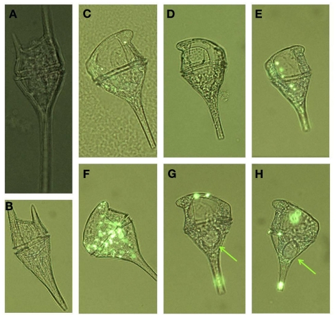
Figure 9.
Micrographs of different forms of Ceratium cells encountered from the red tide incubator region of Monterey Bay showing (A) C. lineatum, (B) C. furca, (C–H) different morphologies of C. balechii. Green arrows in (G,H) indicate the location of feeding vacuoles that were observed in some individuals. Cells are shown under UV illumination with visible light on, and ELF labeling is visible in some cells as bright green regions.

