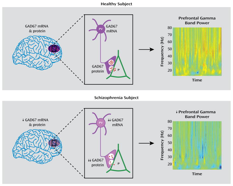
FIGURE 5. Schematic Summary and Predicted Functional Consequences of Lower GAD67 mRNA and Protein Levels in the Dorsolateral Prefrontal Cortex of Subjects With Schizophreniaa.
aIn healthy subjects, normal tissue levels of GAD67 mRNA and protein in the dorsolateral prefrontal cortex (purple oval in top panel, left) are associated with normal levels of GAD67 mRNA and protein in parvalbumin (PV)-containing cell bodies and axon terminals, respectively (top panel, center), and normal amounts of GABA (red dots) release from synaptic vesicles onto pyramidal (P) neurons, providing the inhibitory inputs required for normal prefrontal gamma band power as measured by EEG during cognitive control tasks (top panel, right). In schizophrenia, modest reductions in tissue levels of GAD67 mRNA and protein (bottom panel, left) are associated with pronounced reductions of GAD67 mRNA and protein in PV neurons and axon terminals, respectively (bottom panel, center). The predicted reduction in GABA release from synaptic vesicles leads to the lower gamma band power present over the dorsolateral prefrontal cortex during cognitive control tasks (bottom panel, right). Warmer colors in the heat maps of gamma band power indicate higher power, and cooler colors indicate lower power. Heat maps are from Cho et al. (8). Copyright 2006, Proceedings of the National Academy of Sciences.

