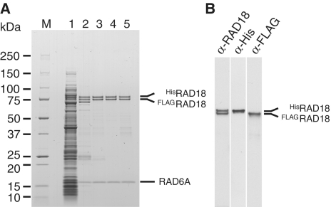
Figure 1.
Purification of the RAD6A–HisRAD18–FLAGRAD18 complex. (A) Pooled fractions eluted from respective columns were analyzed by SDS–PAGE followed by staining with CBB. Lane 1, cell lysate; lane 2, Ni-chelating column; lane 3, heparin column; lane 4, gel-filtration column; lane 5, anti-FLAG affinity column. Molecular masses of each marker (lane M) are shown to the left of the gel. (B) Western analysis of the pooled fraction eluted from the gel filtration column. Membranes were probed with the indicated antibodies.

