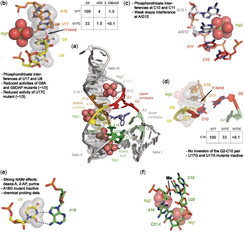
Figure 5.
Visualization of structural features of the Diels–Alderase ribozyme. (a) Overview of the structure. Nucleotides belonging to the ‘spine’ are represented as semi-transparent gray spheres through all panels of this figure. (b) Cross-strand junction stabilized by magnesium ion Mg5 and one H-bond. Inset: krel values of selected atomic mutants. (c) Coordination of magnesium ion Mg3. (d) Importance of the U17–C10 H-bond with atomic mutagenesis data. (e) The reverse Hoogsteen base pair U8–A18 with supporting data. (f) Sharp turn involving nucleotides 18–21.4. The two arrows indicate positions where the addition of a CH3 group interferes with activity.

