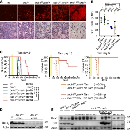
Figure 3.
Conditional deletion of mcl-1 causes regression of AML in tumor-burdened mice. (A) Histological examination of bone marrow from mice burdened with MLL-ENL-induced AML of the indicated genotypes. The top panels show apoptosis of cells (detected by TUNEL staining) after 3 d of treatment with tamoxifen. The bottom panels show the presence of leukemic blasts after 5 d of treatment with tamoxifen. (B) Symptomatic (elevated leukocyte counts, thrombocytopenia, and anemia) mice bearing MLL-ENL-induced AML of the indicated genotypes were treated for 5 d with tamoxifen. Shown are the total numbers of GFP+ leukemic cells collected from two femora and two tibiae on the sixth day. (*) P < 0.05; (**) P < 0.01; (***) P < 0.001. (C) Survival of AML-burdened mice that were treated with tamoxifen (treatment window indicated by yellow bar) 21 d (left panel) 10 d (middle panel) or 5 d (right panel) after transplantation with MLL-ENL transformed AML cells of the indicated genotypes. Untreated mice (dotted lines) from the same cohort treated with tamoxifen at 10 d or 5 d all developed disease, thus confirming the presence of AML at that time pointl (*) P < 0.05; (**) P < 0.01; (***) P < 0.001. (D,E) Western blot analysis to detect Bcl-xL (D) or Mcl-1 and CreER proteins (E) in leukemic cells of the indicated genotypes from mice that relapsed with AML after tamoxifen treatment.

