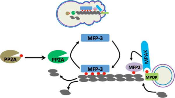
Figure 2.

Schematic diagram depicting the motility machinery of Ascaris sperm. (Top) Cartoon of spermatozoan shows the in vivo location of the proteins participating in the assembly and disassembly of MSP fibers. The outer and inner leaflets of plasma membrane are represented as blue line and violet line, respectively. (Bottom) The MSP dimer (grey) constitutes the building block of MSP fiber. The MSP fiber grown in vitro is composed of two parts: vesicular part and fibrous part. Note that the plasma membrane of the vesicle that supports the assembly of MSP fiber is turned `inside out'. Unknown tyrosin kinase activates MPOP at the leading edge. Phospho-MPOP recruits MPAK which in turn phosphorylates MFP2. Phospho-MFP2 increases the rate of MSP fiber assembly. Unknown kinase phosphorylates cytosolic protein, MFP3 at multiple sites. Phospho-MFP3 binds with MFP3 to stabilize MSP fibers. The Ser/Thr phosphatase, PP2A is localized near the cell body which gets activated upon dephosphorylation by unknown phosphatase. Active PP2A dephosphorylates phospho-MFP3 and dephosphorylated MFP3 detaches from MSP fibers, thereby favoring the disassembly of MSP fibers near the cell body. Phospho group appended to the proteins are depicted as small red circle.
