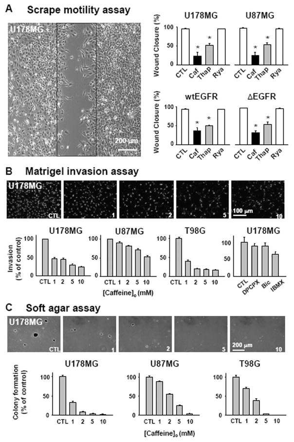
Fig. 2. Caffeine slows motility, invasion, and colony formation of glioblastoma cells.
A. Monolayers of glioblastoma cells were wounded by a scrape (black box) and treated with 10 mM caffeine (Caf), 1 μM thapsigargin (Thap), or 10 μM ryanodine (Rya). All error bars represent SEM. (*p <0.01, ANOVA with Newman-Keuls post hoc.). B. (Top) representative pictures of DAPI labeled cells that invaded through 8 μm holes in the Matrigel inserts in the presence of indicated caffeine concentration. (Bottom) percentage of invaded cells respect to the control condition. Similar experiment was done with 800nM DPCPX, 10 μM Bicuculline, 100 μM IBMX and % of invasion was plotted (last panel). C. Caffeine effect on the anchorage-independent growth of glioblastoma cells in vitro was tested. (Top) representative photographs of colonies grown in indicated caffeine concentration. (Bottom) percentage of number of colony normalized to the control condition.

