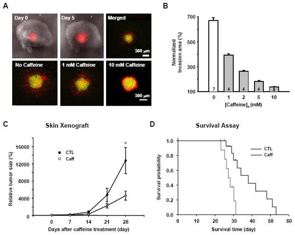
Fig. 6. Caffeine inhibits invasion and increases survival rate.
A. DiI-stained U178MG cells were placed on the surface of slices in the absence or presence of caffeine (0~10 mM) 6 days after slice preparation. The first two merged DIC and red fluorescence images show the hippocampal brain slice and DiI-stained U178MG cells. After 1 h (Green) and 120 h (Red), movement of the glioblastoma cells in the slices was detected with an inverted confocal laser scanning microscope. B. Invasion area (%) was calculated from the formula, Area of DiI-stained cells at 120 h/Area of DiI-stained cells at 1h) × 100. C. Effect of caffeine on the growth of U87MG cells in vivo skin xenograft model (*p<0.01). D. Kaplan-Meier survival curves of nude mouse bearing intracranial U87MG tumors. Log rank, p=0.001, control versus caffeine.

