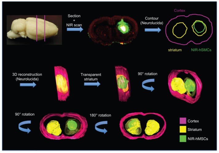
Figure 6.
Three-dimensional reconstruction. Coronal sections were scanned and images imported into the Neurolucida software; contour of whole brain, striata, and transplanted cells was performed and partial three-dimensional images of NIR815 human mesenchymal stem cells within the lesioned striatum were obtained.
Abbreviations: NIR, near-infrared; hMSCs, human mesenchymal stem cells; 3D, three-dimensional.

