Fig. 1.
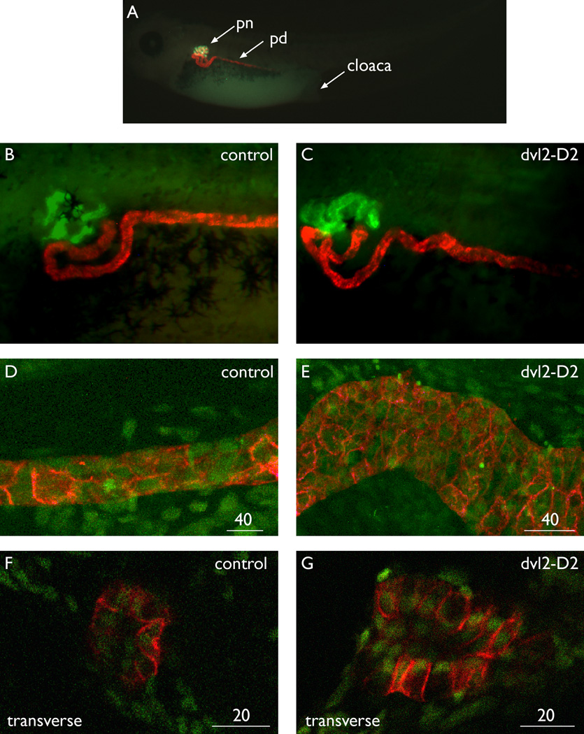
Dominant-negative dvl2 generates Wolffian ducts that are abnormally shaped, wider, and contain more cells than control ducts. Staining details for each technique are available in the Experimental Procedures section. A: Control embryo stained for proximal tubules in green (3G8) and the Wolffian duct in red (4A6). B: Lateral view of control stained for proximal tubules with ECL (green) and 4A6 (red). C: Dvl2-D2 mRNA-injected pronephros. The Wolffian duct is an abnormal serpentine shape. D: Confocal maximal projection of a control duct, stained with 4A6 (red) to highlight the pronephros and counterstained with sytox (green) for nuclei. E: Confocal maximal projection of dvl2-D2 mRNA-injected pronephros, stained as described in D. The duct is wider and has more cells than controls. F,G: Control and dvl2-D2 mRNA-injected ducts stained with 4A6 (red) and sytox (green) then viewed in optical transverse sections. The dvl2-D2 duct contains more cells, but both have an open lumen and normal epithelialization. pn, pronephric nephron proximal domain; pd, pronephric distal tubule and duct; cl, cloaca. Panels A through E show lateral views, while F and G illustrate transverse views. Scale bar = 40 microns in D,E, 20 microns in F,G.

