Figure 3.
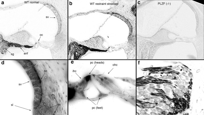
Intense PLZF immunoreactivity is observed in the auditory nerve, organ of Corti, and stria vascularis. a–c, PLZF immunoreactivity from mice at rest (a) or following exposure to 12 h of restraint stress (b) showed similar patterns of labeling, though labeling was more intense following restraint stress. No labeling in the luxoid [PLZF (−/−)] mouse (c) indicates specificity of the antibody. Sections in a–c were immunoreacted together and images were taken at identical exposure levels. d–f, Higher-power images of the lateral wall (d), organ of Corti (e), and spiral ganglion (f) are presented. Strongest label is observed in the auditory nerve, in heads of the pillar cells (pc), and in basal cells (bc) of the stria vascularis (sv), but label is widely distributed throughout the cochlea, including: sv, fibrocytes of the spiral ligament (sl), and blood vessels (bv); the phalangeal processes of Dieter's cells, inner hair cells (ihc), and outer hair cells (ohc) of the organ of Corti (oc) (see also Nagy et al., 2005); and neuronal cell bodies of the spiral ganglion neuron, where punctate label in the cytoplasm is evident. Observations were made on both cochleae from 6 wild-type and 4 homozygous mutant mice.

