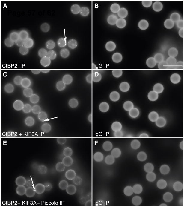
Figure 2. Affinity purified ribbons attached to magnetic beads.
Antibodies for proteins known to be in the ribbon were coupled to magnetic beads. The results were optimized by visualizing the precipitated ribbons with immunolabeling for CtBP2/Ribeye. Ribbons could be pulled down with antibodies for CtBP2 (corresponding to the B domain of Ribeye isoform), KIF3A, and Piccolo. The quantity of ribbons pulled down with CtBP2/Ribeye was not increased by adding antibodies to other ribbon proteins such as Piccolo and/or KIF3A (A, C, E). As a control for nonspecific binding, we used mouse IgG (B, D and F). The beads exhibit a degree of autofluorescence. The bright dots (indicated by arrows) in A, C, and E are partially broken ribbons. Due to the spherical structure of the beads, only the periphery is in focus at these pictures. In reality the entire bead is covered with ribbons. Scale bar (in B) 10 μm.

