Fig. 3.
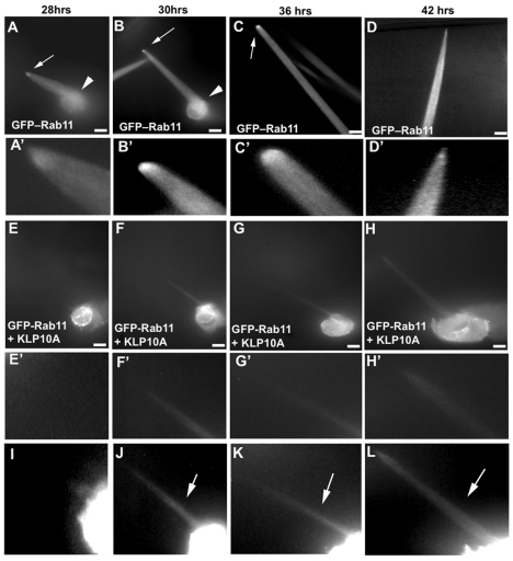
Rab11 intracellular transport is microtubule dependent. Time-lapse imaging of bristles expressing GFP-Rab11 and Klp10A under neur-Gal4 as compared with control bristles expressing only GFP-Rab11. (A-C) GFP-Rab11 localization in a wild-type bristle cell during its growth phase. Note the tip enrichment of Rab11 (arrows). Note the cell body GFP-Rab11 (arrowheads). (D) GFP-Rab11 localization in a wild-type bristle cell near the end of its growth phase. Note the decreased tip enrichment. (A′-D′) Magnified images of the bristle tip from A-D, respectively. (E-G) GFP-Rab11 localization in a bristle expressing Klp10A during its growth phase (starting ∼24 hours APF). (H) GFP-Rab11 localization in a bristle expressing Klp10A after its growth phase. (E′-H′) Magnified images of the bristle tip from E-H, respectively. (I-L) Highly enhanced versions of images E-H that allow the bristle shaft structure (arrows) to be seen. Images are of a single bristle tracked in each set. Images were taken at 60× magnification and represent a projection of optical sections. In control images C and D, the entire length of the bristle cell is not shown. Scale bars: 10 μm.

