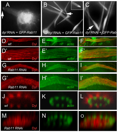
Fig. 6.
Dyl and Rab11 localization in bristles. (A-C) GFP-Rab11 localization in bristle expressing dyl RNAi at (A) 29 hours, (B) 40 hours and (C) 56 hours APF at 29°C. Arrows in A and B indicate tip accumulation of GFP-Rab11 (magnified in inset in B). Arrow in C shows the accumulation of GFP-Rab11 in a blebbing region of a bristle. (D-O) Dyl localization in wild-type and Rab11 mutant bristles visualized by staining with anti-Dyl antibody. Localization of (D) Dyl and (E) F-actin in wild-type bristles. (F) Merge of D and E. (D′-F′) Magnifications of D-F, respectively. (G,H) Localization of Dyl (G) and F-actin (H) in Rab11 kd bristles. (I) Merge of G and H. (G′-I′) Magnifications of G-I, respectively. (J-O) z projections showing Dyl (J,M) and F-actin (K,N) localization around the circumference of wild-type (J,K; merge in L) and Rab11 kd (M,N; merge in O) bristles.

