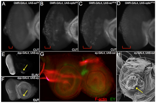
Fig. 5.
Regulatory relationship between Eyeless, Sine oculis and Cut in the eye-antennal disc. (A-G) Confocal images of third instar eye-antennal discs. Brackets in A-D mark the zones of ct-positive cone cells. (H) Scanning electron microscopy of an adult head. Arrows indicate a partial antennal segment in the place of the eye.

