Figure 1.
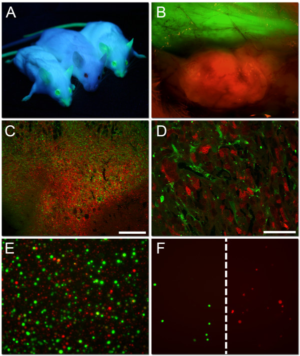
dsRed transfected 4T1 mammary tumor in eGFP mice. A) Two NOD/SCID mice expressing enhanced green fluorescent protein (eGFP) under UV-illumination flanking one plain NOD/SCID mouse from the non-transgenic parental line. B) An in situ picture of a 4T1 dsRed tumor growing subcutaneously in the NOD/SCID eGFP expressing mouse after removing the skin flap (×40 magnification). C and D) Representative confocal microscopy pictures of a 4T1 mammary control tumor, with dsRed expressing tumor cells, as well as infiltrating eGFP expressing host cells. Scale bars indicate 250 μm (C) and 50 μm (D). E) The dissociated 4T1 mammary tumor showing single eGFP expressing host cells together with single dsRed transfected tumor cells (×100 magnification). F) eGFP expressing stromal cells and dsRed expressing 4T1 tumor cells after Fluorescence activated cell sorting (FACS), verify successful separation.

