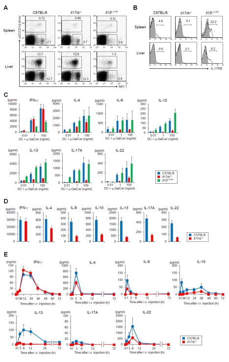
Figure 1. Function of iNKT cells in the spleen and liver from Il17rb −/− and Il15 L117P mice.
(A) FACS profile of spleen and liver mononuclear cells in WT, Il17rb −/− and Il15 L117P mice on a B6 background. Numbers are percentage of gated cells. α-GalCer/CD1d dimer+ iNKT cells and α-GalCer/CD1d dimer− NK1.1+ NK cells were slightly decreased in Il17rb −/− mice and markedly reduced in Il15 L117P mice. (B) IL-17RB expression in spleen and liver iNKT cells of WT B6, Il17rb −/− and Il15 L117P mice. Shaded profiles in the histograms indicate the background staining with isotype matched control mAb. (C, D) In vitro cytokine production by spleen iNKT cells from Il17rb −/− and Il15 L117P mice (C) and by liver iNKT cells from Il17rb −/− mice (D). Sorted iNKT cells (5×10⁴/100 µL) from spleen and liver of WT B6 and Il17rb −/− mice were co-cultured with BM-DCs (5×10³/100 µL) for 48 h in the presence of the indicated doses of α-GalCer. The Il17rb −/− iNKT cells produced IFN-γ at levels equivalent to WT, while TH2 and TH17 cytokine production, except for IL-4, were severely impaired. (E) iNKT cell-dependent cytokine production in WT B6 and Il17rb −/− mice in vivo. α-GalCer (2 µg) was i.v. injected and the levels of cytokines in serum were analyzed at the indicated time points. The serum IFN-γ levels were similar in both mice, whereas production of TH2 and TH17 cytokines, except for IL-4, was significantly reduced in the Il17rb −/− mice. Cytokines were measured by ELISA or a cytometric bead array system at the indicated time points. Data are mean ± SDs from three mice and repeated three times with similar results.

