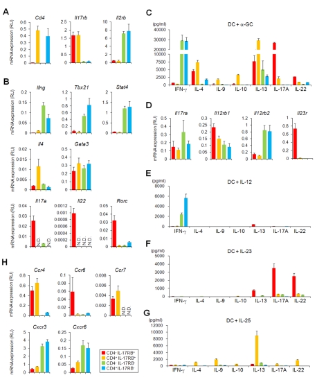
Figure 3. Differential gene expression and cytokine production among thymic iNKT cell subtypes from B6 mice.
(A, B, D, H) Quantitative PCR analysis of thymic iNKT subtypes. Thymic iNKT cells further divided into four subtypes based on the expression of CD4 and IL-17RB (red, CD4− IL-17RB+; orange, CD4+ IL-17RB+; blue, CD4− IL-17RB−; green, CD4+ IL-17RB−). One representative out of three experiments is shown (mean ± SEM). (A) The purity of the sorted cells was confirmed by the relative Il17rb and Cd4 mRNA expression levels in the respective subtypes. Il2rb ( = Cd122) expression was restricted to CD4− and CD4+, IL-17RB− iNKT cells. (B) Expression of TH1/TH2/TH17 related genes. TH1 related: Ifng, Tbx21 and Stat4, TH2 related; Il4 and Gata3, and TH17 related: Il17a, Il22 and Rorc transcripts were analyzed. (D) Expression of cytokine receptor genes. Receptor for IL-12, IL-23, and IL-25 were analyzed. The component chains of the various receptors are IL-12 receptor: IL-12Rβ2/IL-12Rβ1; IL-23 receptor: IL-23R/IL-12Rβ1; IL-25 receptor: IL-17RB/IL-17RA. (H) Expression of chemokine receptor genes. Ccr4, Ccr6, Ccr7, Cxcr3, and Cxcr6. (C, E, F, G) In vitro cytokine production by thymic iNKT cell subtypes (red, CD4− IL-17RB+; orange, CD4+ IL-17RB+; blue, CD4− IL-17RB−; green, CD4+ IL-17RB−). Sorted thymic iNKT subtypes (5×10⁴ cells/100 µL) were co-cultured with BM-DCs (5×10³/100 µL) for 48 h in the presence of α-GalCer (100 ng/µL) (C), IL-12 (10 ng/µL) (E), IL-23 (10 ng/µL) (F), or IL-25 (10 ng/µL) (G). Levels of IFN-γ, IL-4, IL-9, IL-10, IL-13, IL-17A, and IL-22 were analyzed. The data are representative of three independent experiments (mean ± SEM).

