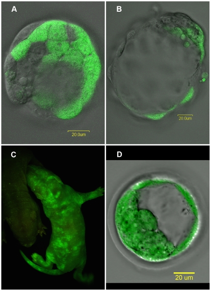
Figure 1. Production of rat chimeras.
Rat chimeras were produced by morulae aggregation where one morula is from a transgenic eGFP lineage and the other is of wild type lineage. (A) These distinct cell lineages can be seen in the embryos before implantation. (B) Further culturing of these embryos reveals the labeled cells integrating into the inner cell mass. (C) A newborn chimera pup, right half of photograph, can be easily distinguished from a non-chimeric littermate, barely seen in the left half of the photograph, when visualized under blue light. (D) A transgenic eGFP blastocyst. Some areas of the transmitted image, where the light isn't restricted by the confocal aperture around the zona pelucida and the periphery of the blastocyst are out of focus which doesn't occur in the fluorescent image where all cells in the inner cell mass as well as the trophectoderm are clearly labeled with eGFP. Scale bars in (A), (B) and (D) are 20 µm.

