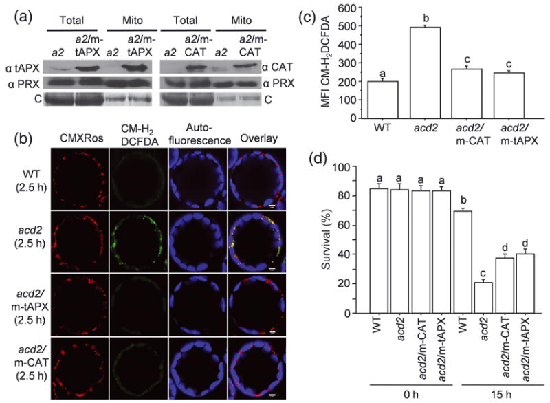
Figure 1. Targeting anti-oxidant enzymes to mitochondria increases the survival of acd2 cells.

(a) Total proteins and mitochondrial proteins were isolated from acd2 (a2), a2/m-tAPX and a2/m-CAT plants and tAPX, catalase (CAT) and mitochondrial marker peroxiredoxin (PRX) II F proteins were detected by Western blot analysis. tAPX and CAT were targeted to mitochondria in the a2/m-tAPX and a2/m-CAT plants, respectively. C= Coomassie staining of the membranes. This experiment was done three times with similar results.
(b) Protoplasts isolated from 20 d-old plants were exposed to light for 2.5 h and double-stained with CM-H2DCFDA to detect H2O2 and CMXRos to mark mitochondria. Fifty to sixty protoplasts were examined under laser scanning confocal microscope (LSCM) and representative protoplasts are shown. Staining with CMXRos validated that CM-H2DCFDA-positive organelles were mitochondria. This experiment was done three times with similar results.
(c, d) The mean fluorescence intensity (MFI) of CM-H2DCFDA-stained protoplasts determined using flow cytometry (c). Protoplasts were exposed to light for 15 h and the percentage (%) of surviving cells was determined by FDA staining (d). Results are from a single analysis, representative of three independent experiments that showed similar results. The error bars represent SD (n=3). Letters indicate different values using Fisher’s Protected Least Significant Difference (PLSD) test (P < 0.004).
