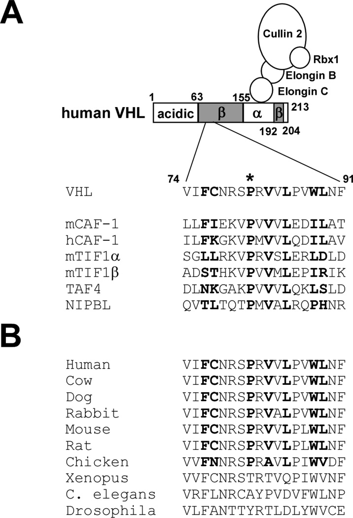
Figure 1. HP1-binding motif in VHL.
(A) The PXVXL motif in human VHL and its alignment with other PXVXL-containing proteins. Bold residues in mCAF-1 are involved in hydrophobic interaction with HP1 chromo shadow domain. Proline 81 (asterisk) is mutated to serine in renal carcinoma-associated VHL P81S mutant.
(B) Sequence alignment of the PXVXL motif in VHL from different species.

