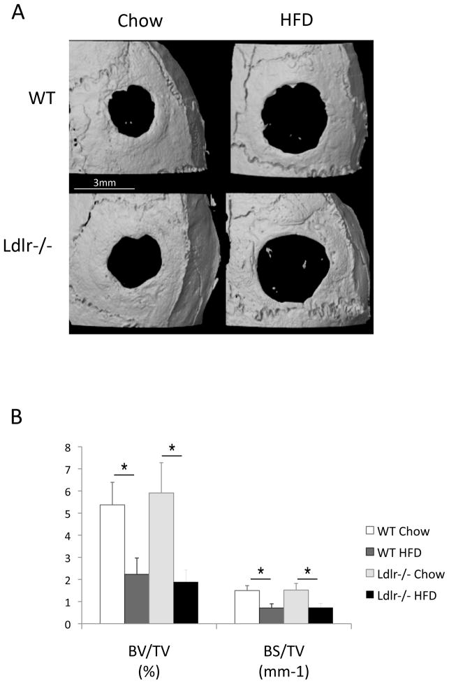
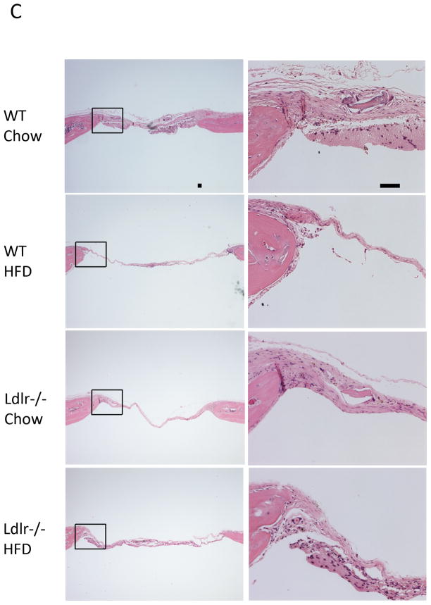
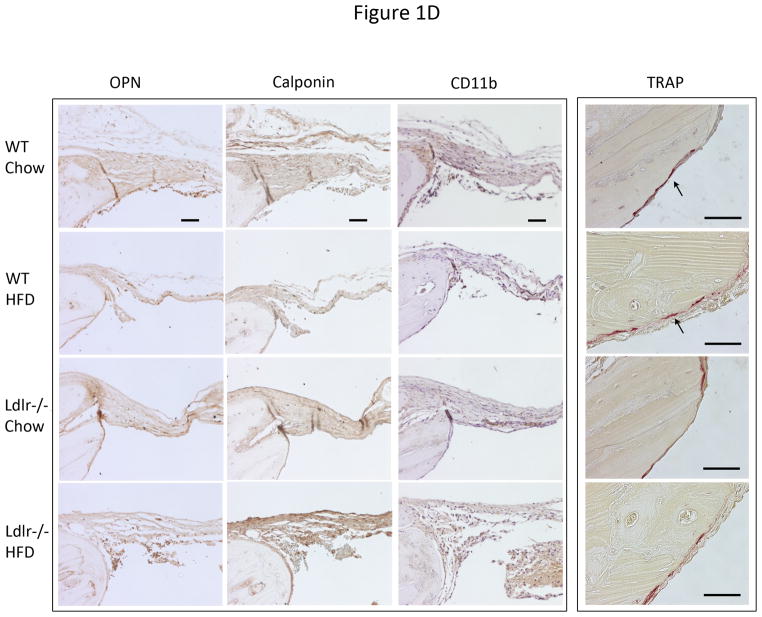
Figure 1. Effects of the HF diet on bone regeneration.
(A) Three-dimensional microCT images of cranial defects in WT and Ldlr−/− mice, with or without the HF diet (HFD). (B) MicroCT analysis of cranial bone volume (BV/TV) and surface (BS/TV). *p < 0.05. (C) Hematoxylin and eosin analysis of cranial bones. Granulation tissue (arrows), which is distinguished from the regenerative fibrous material by its hypercellularity, lack of fibers, and lack of connection with the adjacent bone, was also present in some sections. (D) Immunohistochemical staining for osteopontin (OPN), calponin, CD11b and histochemical staining for TRAP. CD11b was counterstained with hematoxylin. Scale bars -50 μm.


