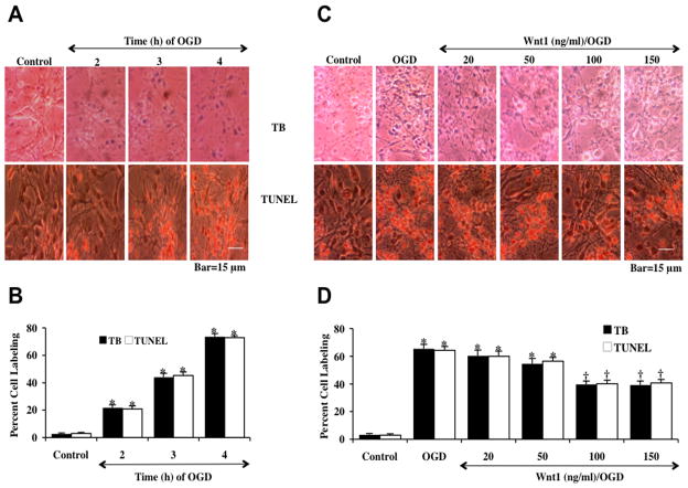
Fig. 1. Wnt1 prevents neuronal cell injury against oxygen-glucose deprivation (OGD).
(A) Primary hippocampal neurons were exposed to progressive durations of OGD of 2, 3 and 4 hours and cell injury was determined by trypan blue (TB) dye exclusion and TUNEL 24 hours after OGD. Representative images illustrate that OGD led to progressive neuronal cell injury with increased exposure time of OGD. In all cases, control = untreated neuronal cells. (B) Quantitative analysis reveals that the percent cell trypan blue staining and apoptotic DNA fragmentation was significantly increased 24 hours following OGD (*p<0.01 vs. control). Each data point represents the mean and SEM from 6 experiments. (C) Increasing concentrations (20, 50, 100, and 150 ng/ml) of Wnt1 protein was applied to neuronal cultures 1 hour prior to a 3 hour period of OGD and cell injury was determined by trypan blue (TB) dye exclusion and TUNEL 24 hours following OGD. Wnt1 at the concentrations of 100 ng/ml and 150 ng/ml significantly reduced neuronal cell labeling of trypan blue (TB) and TUNEL 24 hours after OGD. (D) Quantitative analysis showed that Wnt1 (100 ng/ml and 150 ng/ml) administered 1 hour prior to OGD significantly decreased the percent cell labeling of trypan blue and percent DNA fragmentation 24 hours following OGD (*p<0.01 vs. control; †p < 0.01 vs. OGD). Each data point represents the mean and SEM from 6 experiments.

