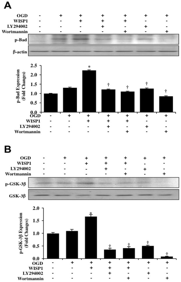
Fig. 4. WISP1 promotes Bad and GSK-3β phosphorylation through PI 3-K and Akt1 pathways during OGD.
(A) Equal amounts of neuronal protein extracts (50 μg/lane) were immunoblotted with antibody against phosphorylated Bad (p-Bad) 3 hour following a 3 hour period of OGD. Application of WISP1 (10 ng/ml) 1 hour prior to OGD significantly increased p-Bad expression compared with OGD treated alone. Combined application of the specific PI 3-K inhibitors wortmannin (0.5 μM) or LY294002 (10 μM) prevented WISP1 phosphorylation of Bad 3 hours following OGD (*p<0.01 vs. OGD; †P <0.01 vs. WISP1/OGD). (B) Equal amounts of neuronal protein extracts (50 μg/lane) were immunoblotted with antibody against phosphorylated GSK-3β (p-GSK-3β) 3 hours following a 3 hour period of OGD. WISP1 (10 ng/ml) administered 1 hour prior to OGD significantly increased p-GSK-3β expression compared with OGD alone. Combined application of wortmannin (0.5 μM) or LY294002 (10 μM) prevented WISP1 phosphorylation of GSK-3β following OGD (*p<0.01 vs. OGD; †P <0.01 vs. WISP1/OGD). In A and B, quantitative analysis of western blots from 3 experiments was performed using the public domain NIH Image program (developed at the US National Institutes of Health and available on the Internet at http://rsb.info.nih.gov/nih-image/).

