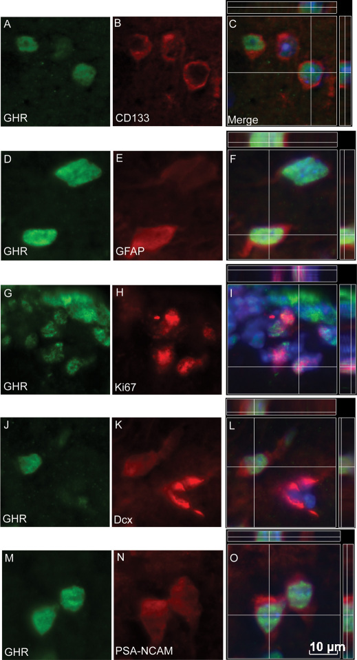
Figure 1. Distribution and phenotype of GHR+ve cells in the adult SVZ.
GHR+ve cells were detected scattered throughout the SVZ surrounding the lateral ventricle. These cells co-expressed the stem cell markers CD133 (A–C) and GFAP (D–F), as well as the cell proliferation marker Ki67 (G–I). GHR+ve cells were also detected in the dorsolateral corner of the lateral ventricle localized with Dcx (J–L) and PSA-NCAM (M–O). DAPI was used to label the nucleus (Blue in merged images) Omission of primary antibody resulted in loss of immunoreaction. Scale bar: = 10 μm.

