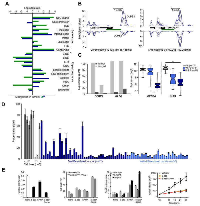
Figure 3. Core promoter methylation in mediators of adipogenesis.
A. Patterns of somatic methylation enriched or depleted (measured by log odds ratio relative to background sequence in the human genome) among 19 sequence contexts including the canonical gene cassette. Tumor-specific increases and decreases of methylation are green and blue, respectively. B. Methylation of CEBPA and KLF4 is increased in tumors (blue), but not their matched normal adipose tissues (gray) in upstream regions or in regions spanning adjacent CpG islands (green). Methylation signal for each tumor is plotted for both the positive and negative strands (dotted and dashed respectively) and combined into total signal (solid) for each sample. Also indicated is the distance between the peak of methylation in both promoters and their respective transcription starts sites. C. CEBPA and KLF4 expression, inferred from RNA sequencing in the methylated samples (normalized rpkM) indicates their reduction in DLPS1 and DLPS2 compared to their matched normal adipose tissues (left panel). On the right, expression measured by microarray in a cohort of 115 tissues (as shown; starred, p-values < 10−12, ANOVA). D. CEBPA promoter methylation status (average percent methylated, two biological replicates assessed by bisulfite pyrosequencing, error bars represent standard deviation) in 8 cell lines (dark and light gray are dedifferentiated and well-differentiated cell lines, respectively) and 72 tumors indicated that CEBPA methylation is high in cell lines and a subset of DLPS, but absent from WLPS tumors. E. Proliferation of DDLS8817 dedifferentiated liposarcoma cells after treatment with 5-aza, SAHA, or the combination of both (left; mean ± propagated error). Proliferation levels are shown relative to untreated cells. The percentage of apoptotic cells is shown (middle, left; measured by annexin V and 7-AAD staining, mean percent positive ± standard deviation) in untreated and drug-treated DDLPS8817 cells. Expression of early, intermediate, and late markers of differentiation (perilipin, FABP4, and adipsin respectively) were measured by RT-PCR (mean ± standard deviation) in the presence of drug and shown relative to the level in untreated cells (middle, right). The growth of DLPS tumors in mice (DDLS8817 xenografts) treated with 5-aza (decitabine), 5-aza plus SAHA, or vehicle (right; mean tumor volume ± standard error of the mean, n = 5 mice/group) was also analyzed.

