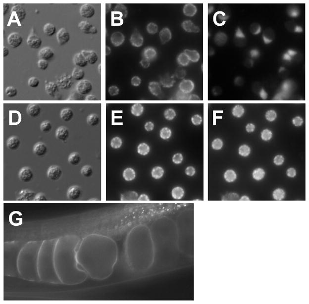
Figure 8.
Examples of sperm and oocyte protein localization. (A–C) Localization of 1CB4 and SPE-9. (A) Nomarski DIC micrograph of mature spermatozoa. (B) Localization of 1CB4 to MOs. (C) Localization of SPE-9 to pseudopods. (D–F) Co-localization of 1CB4 and SPE-38 in spermatids. (D) Nomarski DIC micrograph of spermatids. (E) Localization of 1CB4 to MOs. (F) Localization of SPE-38 to MOs. Note the identical distribution of staining in panels E and F. (G) GFP fluorescence of a GFP:EGG-1 fusion protein in oocytes.

