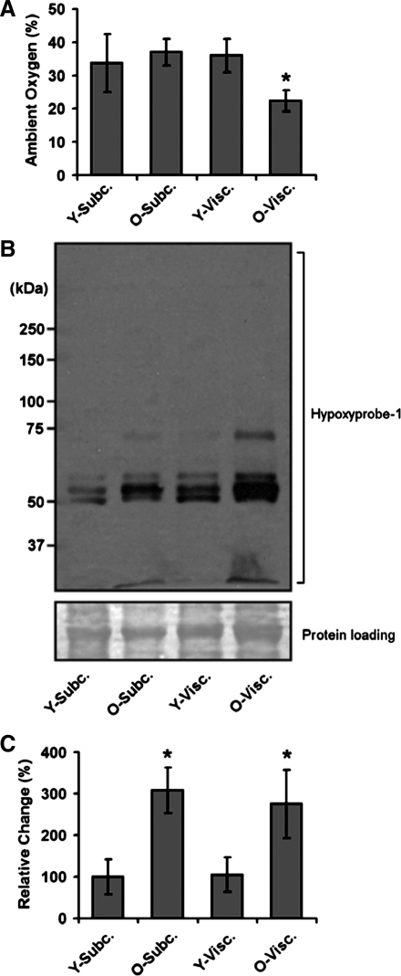
Fig. 2.
Aging induces hypoxia in adipose tissue. A: adult (6-mo- and 26-mo-old) mice were anesthetized, and levels of oxygen measured directly within visceral (epididymal) and subcutaneous (inguinal) fat depots using insertion of an oxygen electrode into the specific depot. B: hypoxia was measured in visceral and subcutaneous fat depots using the Hypoxyprobe method, and results from multiple experiments were quantified (C). See materials and methods for experimental details. Data are expressed as means ± SE and represent results from n = 8 for each experimental end point. *P < 0.05 vs. young (Y, 6-mo-old) adult mice.

