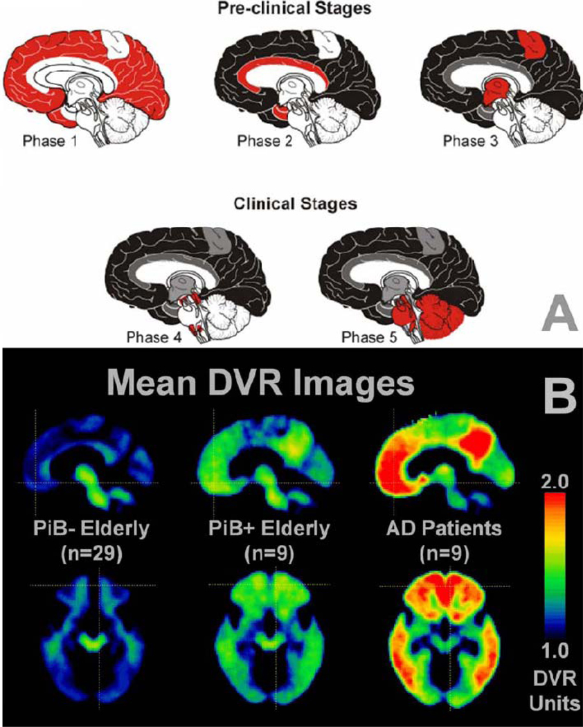
Figure 1. The progression of amyloid deposits as assessed at different stages of AD in (A) post-mortem brain tissue using immunohistochemical techniques against Aβ and (B) in vivo amyloid-PET scans.
(A) Phases (1–5) of postmortem histological appearance of Aβ in clinical AD, as based on [125]. Note that the stages refer to Aβ brain deposition and not necessarily to clinical severity. In Phase 1, Aβ is largely restricted to neocortical brain areas. In phases 2 and 3, when clinical signs of cognitive decline have yet to appear, Aβ deposits are localized throughout the neocortex, allortex, including brain areas such as the medial temporal lobe, and also begin to occur in the motor cortex and subcortical brain structures. In the clinical stages of AD (phase 4 & 5), Aβ deposits are observed globally in the brain including the brain stem and the cerebellum in the final stage of the disease (phase 5). Key: white, no Aβ deposits; red, novel Aβ deposition; grey, Aβ deposits that were missing in phase 1; black, Aβ deposits that were present already in phase 1 (B) A similar spatial pattern of progression of Aβ deposition is observed when using PiB-PET in cognitively normal elderly controls who appear to be Aβ-free (PiB−), cognitively normal elderly controls who appear to be in the early stages of Aβ deposition (PiB+) and in symptomatic AD patients [18]. Mean sagittal distribution volume ratio (DVR; cerebellum as reference) images are at the top and transaxial images at the bottom. Reproduced, with permission, from [125] (A) and [18] (B).

