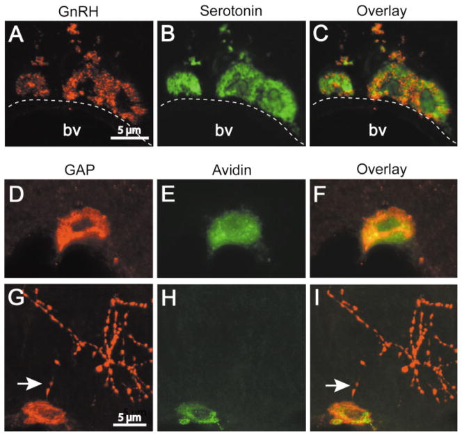
Figure 1.
GnRH-I and GAP immunoreactivity in mast cells in the rat thalamus and median eminence. (A–C) Confocal images (1 μm) of brain mast cells containing GnRH-I- (A) and serotonin- (B) IR. The overlay (C) shows that both labels occur in the same cell but not necessarily in the same mast cell granule. (D–I) Confocal images of GAP (D and G) and avidin (E and H) labeled mast cells in the thalamus and median eminence, respectively. Note that GAP and avidin staining occur in the same mast cell (F and I). GAP, but not avidin, is present in axons in the median eminence (G–I). Note the proximity of GAP positive axons (arrows) to mast cells. D–F and G–I are projections of 6 and 8 images (z axis, 1 μm), respectively. [Color figure can be viewed in the online issue, which is available at www.interscience.wiley.com.]

