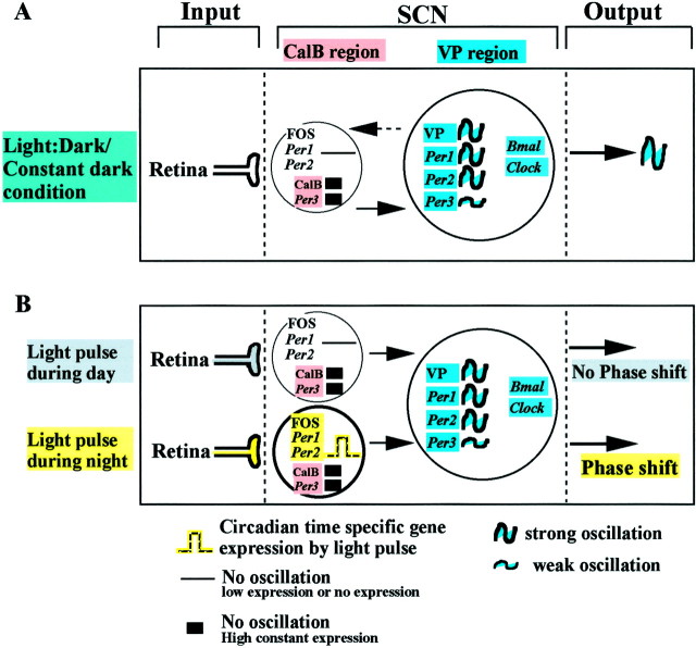
Fig. 4.
Model of rhythmicity and photic entrainment at the tissue level, comprising two functionally different SCN compartments. As is widely accepted, the circadian system has three primary components: input, SCN, and output. A,Model of rhythmicity. In both Light:darkand Constant dark conditions, there are two distinct regions of the SCN showing the same pattern of Per gene expression. In the hamster SCN, these regions can readily be delineated by cells containing CalB and VP respectively, as indicated in Materials and Methods. These two compartments constitute the SCN pacemaker. In the CalB region, rhythmic expression of Per1 andPer2 mRNA are not detectable (SCN,left). Another characteristic of this area is that CalB and Per3 mRNA are highly expressed but not rhythmic. Endogenous rhythmic expression of clock genes (Per1, Per2, Per3, and Bmal1 mRNA) and VP mRNA occurs in a large number of SCN cells restricted to the VP region (SCN, right). B. Model of photic entrainment. Gated light induced Per1 and Per2 mRNA and FOS expression. A light pulse during the day (top) has no effect on the expression of Per1 and Per2mRNA or on FOS. A light pulse during the night (bottom) induces Per1 and Per2 mRNA and FOS expression in the CalB region. Importantly, some genes in this region are not induced by light pulse during the night (Per3, CalB). See Discussion for the proposed mechanism of entrainment.Ant, Anterior; Post, posterior.

