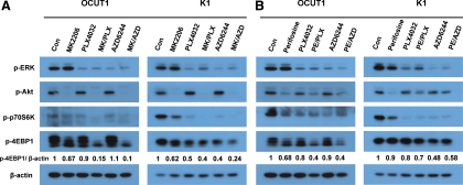
Fig. 2.
Effects of the Akt inhibitors and BRAFV600E/MEK inhibitors, used individually or in combinations, on the MAPK and PI3K/Akt signalings in thyroid cancer cells. A, OCUT1 and K1 cells were treated with 1 μm MK2206, 0.5 μm PLX4032, or 0.2 μm AZD6244, individually or in the indicated combinations (MK/PLX and MK/AZD, respectively) for 24 h, followed by cell lysis in radioimmunoprecipitation assay buffer. Cell lysates were subjected to Western blotting with antibodies against p-ERK, p-Akt, p-p70S6K, p-4EBP1, or β-actin. β-Actin was used for quality control. Con, Control. B, OCUT1 and K1 cells were treated with perifosine (3 μm for OCUT1 cells and 10 μm for K1 cells), 0.5 μm PLX4032, and 0.2 μm AZD6244, individually or in the indicated combinations (PE/PLX and PE/AZD, respectively) for 24 h, followed by Western blotting as described in A. Relative quantification of p-4EBP1, as shown underneath the band, was determined by the ratio of the density of p-4EBP1 of different treatments to control vehicle treatments, which had already been divided by the density of their respective β-actin bands for protein correction.

