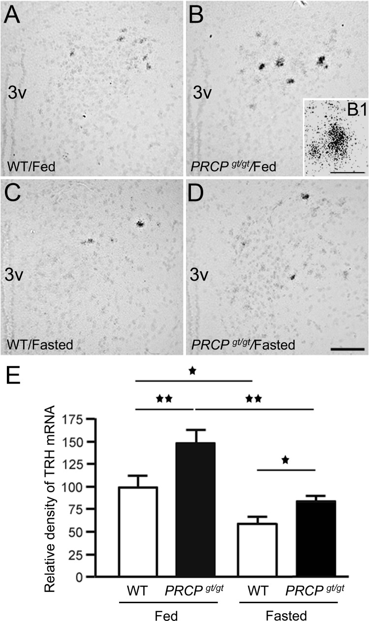
Fig. 4.
Representative micrographs of PVN from WT and PRCPgt/gt mice fed on a regular chow diet (A and B, respectively) and overnight-fasted condition (C and D, respectively) showing the silver grains from radioactive in situ hybridization of TRH mRNA on Nissl-stained background. 3V, Third ventricle. B1, A high power micrograph of a PVN neuron of the fed PRCPgt/gt mouse showing silver grains labeling. E, Results from the analysis of the expression levels of TRH mRNA from each group. TRH mRNA expression levels were measured using ImageJ program and relative density of silver grains was compared between groups. *, P < 0.05; **, P < 0.01. Scale bars, 50 μm (A–D) and 10 μm (B1).

