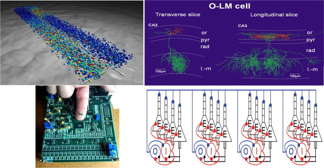
Figure 1.
Top left, Full-scale network simulation of the network coordinating locomotion in the lamprey–intersegmental coordination in a complete spinal cord. This large-scale network model consists of approximately the correct number of excitatory interneurons and commissural inhibitory interneurons (10,000 Hodgkin-Huxley, 5-compartment models) along the entire spinal cord. The model neurons express the different subtypes of ion channels found experimentally. The excitatory synaptic interaction is via AMPA and NMDA receptors and the inhibition via glycine Cl− channels. The wave of activity is transmitted from rostral to caudal, with the left and right sides alternating. Red dots represent actively spiking neurons, yellow ones are depolarized but are not firing, and blue dots are inhibited cells. The network extends from the rostral (bottom right) to the caudal (top left; segment 100) aspect. Segment 50 is indicated with a hatched line, and the perspective is thus compressed caudally. Top right, Biocytin-labeled O-LM interneuron in transverse and longitudinal slices of CA3 hippocampus. Note the longer extension in the longitudinal slice. Bottom right, The network is an abstraction used for numerical simulations to explain how these interneurons, which fire at a theta (4–12 Hz) frequency, can coordinate spatially separated cell assemblies firing at the higher gamma band frequencies. Such analyses permit exploration of emergent properties of interacting neurons and networks of neurons (Tort et al., 2007). Bottom left, A neuromorphic chip (designed by John Arthur, Stanford University, Stanford, CA) that models 1024 pyramidal cells and 256 interneurons in the hippocampus. The chip is housed in a black plastic package and mounted on a printed circuit board. or, Stratum oriens; pyr4, stratum pyramidale; rad, stratum radiation; l-m, stratum lacunosum-moleculare.

