
Fig. 1.
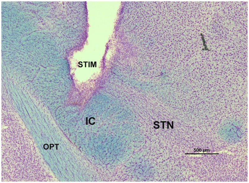
STN stimulator placement. A representative micrograph showing stimulating electrode placement in a coronal section through the rat STN stained via the Kluver-Barrera method. The luxol fast blue component of Kluver-Barrera stains the fiber tracts and the cresyl violet component stains the cell bodies. Therefore, this staining makes the dense cellular swath of the STN immediately dorsal to the internal capsule easily identifiable. Electrode placement was considered to be appropriately targeted to the STN if the tip was within or at the border of the STN within any of the sections examined. STIM = stimulator, IC = internal capsule, OPT = optic tract, STN = subthalamic nucleus. Scale bar = 500μm.