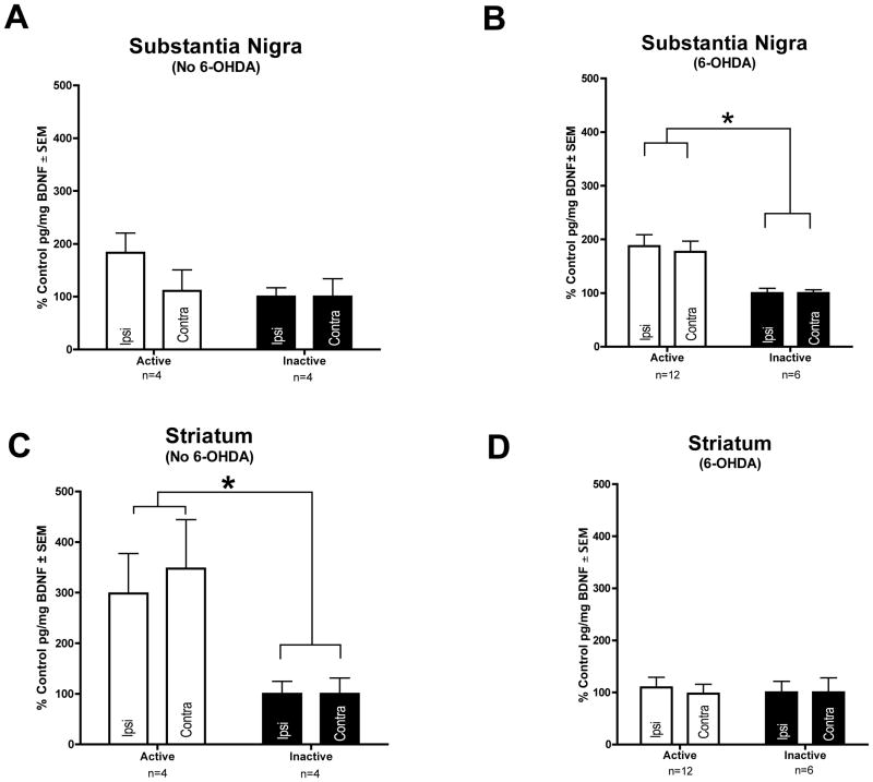
Fig. 4.
Long Term STN DBS increases BDNF protein in the nigrostriatal system. No increase in nigral BDNF protein was seen with ACTIVE stimulation in intact rats (n = 4) (A). In 6-OHDA lesioned animals BDNF protein is bilaterally increased in the substantia nigra of rats receiving ACTIVE stimulation (n = 12, white bars) as compared to INACTIVE stimulator controls (n = 6, black bars) (B, *p < 0.05). BDNF protein was bilaterally increased in the striatum of rats receiving ACTIVE stimulation (n = 4, white bars) as compared to INACTIVE stimulator controls (n = 4, black bars) in intact rats (C, *p < 0.05), but was not increased in the striatum of 6-OHDA lesioned rats (ACTIVE n = 12, INACTIVE n = 6) (D). Values expressed as the mean percent of control ± SEM for each group. The control for the ACTIVE left hemisphere (stimulator and 6-OHDA hemisphere) was the mean BDNF pg/mg protein value of the INACTIVE left hemisphere and the control for the ACTIVE right hemisphere was the mean BDNF pg/mg protein value of the INACTIVE right hemisphere.

