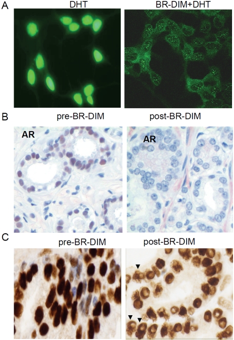
Figure 3.
BR-DIM treatment inhibited AR expression and prevented AR from nuclear translocation. (A) Immunofluorescence staining for androgen receptor (AR) captured through confocal microscopy. LNCaP cells were cultured in RPMI1640 medium with 10% charcoal-stripped serum and kept untreated (strong nuclear staining, left panel) or treated with 25 μM BR-DIM (weaker cytoplasmic staining and nuclear exclusion, right panel) for 24 hours followed by 1 nM DHT treatment for 2 hours. (B) Immunohistochemical staining for AR using human PCa tissue specimen prior to BR-DIM intervention (diagnostic biopsy, left panel) showing strong nuclear AR staining, and after BR-DIM intervention (radical prostatectomy specimen after BR-DIM intervention, right panel) showing negative AR staining in the nuclear compartment. (C) Immunohistochemical staining for AR using human PCa tissue specimen prior to BR-DIM intervention (diagnostic biopsy, left panel) showing dark nuclear AR staining, and after BR-DIM intervention (radical prostatectomy specimen after BR-DIM intervention, right panel) showing reduced AR staining and nuclear exclusion of AR (arrowheads).

