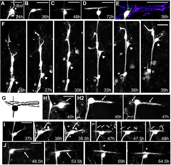
Figure 2.
Development of the fasciculated dbd arbor. (A-D) Representative in vivo confocal z-projections of dbd migration and arbor formation from 24 h to 72 h APF. (E) Initial outgrowth onto immature myoblasts: green, anti-CD8; magenta, phalloidin F-actin. (F) Z-projections from two-photon time-lapse videos showing dbd (arrowhead) and neuron ddaE (e) between 25 h and 39 h APF. (G) Cartoon of the branches within the dbd arbor. (H-J) Time-lapse z-stacks showing four features of dbd branching and elaboration. (H1) and (H2) show posterior growth cone reversal (arrows), and new branches from the soma (asterisks). (I, J) Arbor thickened through adhesion of growth cone filopodia (large arrows), and interstitial filopodia (small arrows), with occasional branch pruning (arrowheads). Scale bar = 30 μm.

