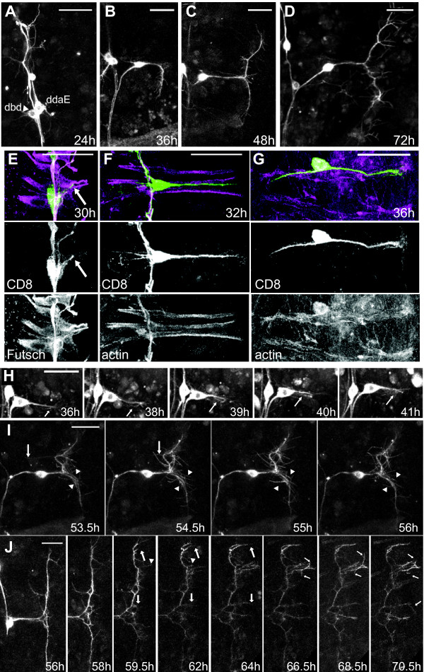
Figure 5.
Elaboration of the BrZ3 ectopic arbor. (A-D) Representative in vivo confocal z-projections of dbd[+BrZ3] at various times during metamorphosis. (E-G) Confocal z-projections of dbd[+BrZ3] at 30, 32, and 36 h APF showing the relationship of the extending dendrite to the underlying myoblasts. Green, anti-CD8; magenta, anti-Futsch (E) or phalloidin F-actin (F, G). The muscle groove in (G) was stretched apart in preparation. Dbd growth cones (arrows). (H-J) Videos of branching in dbd[+BrZ3]. (H) Fasciculation through growth cone reversal (arrows). (I) The rare ectopic branches that attempted to fasciculate were removed (arrowheads), as were branches growing into the next muscle groove (small arrows). (J) Rapid extension of secondary branches (large arrows) followed by limited stabilization of tertiary filopodia (small arrows). Significant branches can be retracted (small arrowheads). All times are hours APF. Scale bar = 30 μm.

