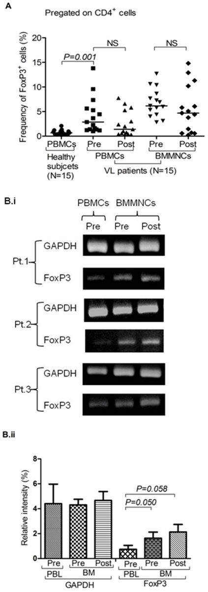
Figure 2. Persistence of Treg cells among visceral leishmaniasis (VL) patients:
A) Frequency of CD4+ FoxP3+ cells after successful therapy: Scatter plot depicting no statistically significant changes in the frequency of FoxP3+ Treg cells after successful therapy (post) in the BM-MNCs and PBMCs of cured VL cases as compared to its pre-treatment levels (pre). Horizontal line in dot plot depicts median value. B) Increased expression of FoxP3 mRNA in BMNCs of patients and their persistence after successful therapy: (i) Gel photograph showing increased expression of FoxP3 mRNA in BM-MNCs as compared to autologous PBMCs. Persistence of FoxP3 mRNA after successful therapy (post) is also shown in pictures. Individual experiments of three patients with their follow up are shown herewith. (ii) Relative density analysis shows increase in FoxP3 mRNA in patient's BMMNCs at pre (Mean±SD, p = 0.050, Mann-Whitney test) and post treatment level (Mean±SD, p = 0.058, Wilcoxon sign rank test). However, GAPDH remain unchanged in all three categories.

