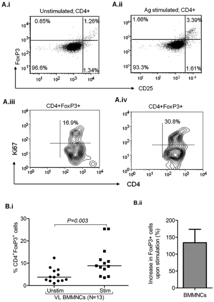
Figure 3. L. donovani antigen driven induction of FoxP3+ cells in BM-MNCs of visceral leishmaniasis (VL) patients: A i–ii.
) Representative plot shows increased frequency of FoxP3+ cells (%) in MNCs upon in vitro stimulation with LD antigen (whole cell lysate, WCL). A iii–iv) Representative plot shows increase in the positivity of Ki67 (an intra nuclear cells proliferating antigen) among gated CD4+FoxP3+ cells upon antigen stimulation (30.8%) as compared to unstimulated cells (16.9%). B i) Scatter plot representing the frequency of Foxp3+ Treg cells in gated CD4+ cells BM-MNCs (n = 13) of VL patients upon in vitro stimulation with LD antigen. Significant increase in the frequency of FoxP3+ Treg cells from BM-MNCs of VL patients occur upon antigen stimulation (p = 0.003, paired t test). Horizontal line in dot plot depicts median value. B ii) Plot shows increase in the frequency of FoxP3+ cells in BMMNCs of VL patients upon stimulation (% increase = (frequency of Treg cell in stimulated culture- unstimulated culture)/frequency of Treg in unstimulated×100).

