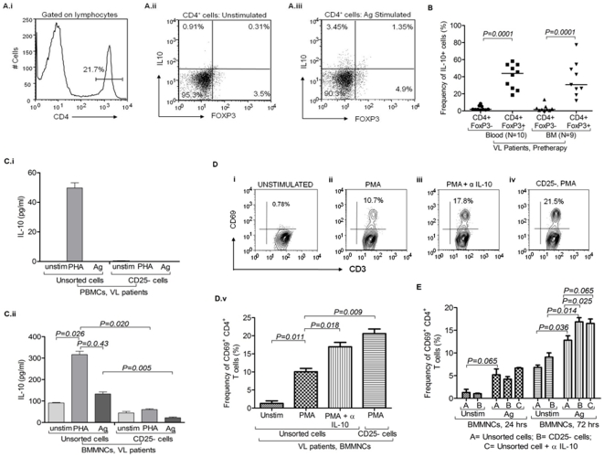
Figure 4. IL-10 production by the Treg cells derived from visceral leishmaniasis (VL) patients:
A) IL-10 producing FoxP3+ cells in BMMNCs of VLs patients before treatment: (i) Histogram plot depicts gating strategy of CD4+ cells. Bi-variant plots show IL-10 production by CD4+FoxP3+ Treg cells and CD4+FoxP3− cells under different in vitro conditions (ii) without stimulation and (iii) with L. donovani antigen stimulation. B) IL-10 production by CD4+FoxP3+ and CD4+FoxP3− cells: Scatter dot plot shows frequency of IL-10 producing cells within gated CD4+FoxP3+ (Treg) and CD4+FoxP3− (Teff) cells in blood as well as in BM of VL patients before anti-Leishmania therapy. Data shows that FoxP3+ cells are one of the important producers of IL-10 along with CD4+FoxP3− cells. Horizontal lines in dot plot depict median value. Significant differences are indicated with p-values using paired t test. C) CD4+CD25+ (FoxP3 enriched) cells are major producer of IL-10 at disease site (BMA): CD25+ cells were magnetically sorted out from (i) PBMCs and (ii) BMMNCs. IL-10 was measured in supernatant of cultured cells (unsorted cells and CD25 depleted cells) stimulated with PHA (mitogen) and L. donovani antigen. IL-10 production was dominantly restricted to CD25+ cells which were enriched with Treg cells. Data are represented in Mean± SD. Significant differences are indicated with p-values using paired t test. D) Effect of IL-10 blocking and depletion of FoxP3+ enriched cells on T cell activation upon polyclonal stimulation: i–v) In vitro PMA stimulation for 24 hrs caused activation of CD4 T cells (CD3+CD8−) derived from BM-MNCs as measured by the expression of CD69 (% positive cells) on them (v; p = 0.011, paired t test). Significant increase in the frequency of CD69+ early activated CD4 (CD3+CD8−) T cells occurred upon PMA stimulation of BM-MNCs for 24 hrs when endogenously produced IL-10 was blocked by monoclonal antibody (v; p = 0.018, paired t test). Similar increase in the frequency of CD69+ CD4 (CD3+CD8−) T cells was also observed when CD4+CD25+ (Treg enriched) were sorted out using MACS sorting kit and CD25− BMMNCs were cultured with PMA for 24 hrs (v; p = 0.009, paired t test). Data are represented in Mean± SD. E) Treg cells and IL-10 suppress L.donovani specific activation of CD4+ T cells derived from pathologic site: LD antigen caused significant increase in the frequency of CD69+CD4+ T cells (p = 0.036, paired t test) after 72 hrs of stimulation. Upon blocking of soluble IL-10 by anti IL-10 ab, the frequency of CD69+CD4+ T cells were increased as compared to antigen alone (p = 0.065, unpaired t test). Upon stimulation of CD25 depleted BMMNCs with antigen for 72 hrs, the frequency of CD69+CD4+ T cells was increased as compared to antigen stimulated unsorted cells (p = 0.025, paired t test) as well as unstimulated CD25 depleted cells (p = 0.014, paired t test). However, in 24 hrs stimulation experiments, unsorted (with/without blocking of IL-10) and CD25 depleted BMMNCs upon stimulation with LD antigen show no significant increase in the frequency of CD69+CD4+ T cells. {A = unsorted cells (with/without ag stimulation), B = CD25 depleted cells (with/without ag stimulation), C = unsorted cells with IL-10 blocking}. Data are represented in Mean± SD.

