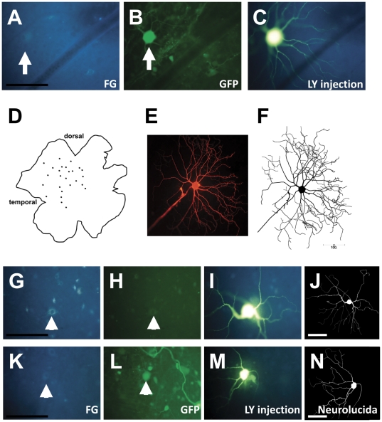
Figure 1. Injection, tracing and identification of individual RGCs from rAAV2-injected retinae.
Photomicrographs and Neurolucida-traced images showing the procedure for labeling and identifying regenerating retinal ganglion cells (RGCs). Regenerating RGCs were first identified based on retrograde labeling (Fluorogold; A), and transduced cells were based on GFP expression (GFP+ RGC is shown in B). After filling the dendrites with Lucifer yellow (C), the RGC was again photographed. This procedure was repeated on 20–50 RGCs per retina (D). The visualization of dendritic architecture was further enhanced with Lucifer yellow immunohistochemistry, and individual cells with complete fills (E) were traced using Neurolucida software. The Neurolucida trace (F) was compared to images of the cells taken immediately after the Lucifer yellow injection (C) to allow each cell to be classified as transduced (GFP+) or as a non-transduced “bystander” neuron (GFP−). G–N: Representative images of RGCs that were retrogradely labeled with Fluorogold (G, K), identified as GFP− (H) or GFP+ (L), injected with Lucifer yellow (I,M) and traced using Neurolucida software (J,N). Scale bars: 100 µm.

