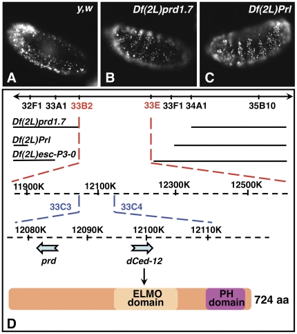
Figure 1. Overlapping deficiencies of the 33B-E genomic region lack clustering of AO-stained apoptotic cells.
In A–C, embryos were aged to stage 13, stained with AO, viewed and imaged with a 20× objective under an epifluorescent Nikon microscope using the red channel, and are presented in a lateral view. A shows a wild-type embryo with a typical pattern of distribution of AO-stained apoptotic cells clusters around the brain lobes and in the posterior end of the embryo. B and C are Df(2L)prd1.7 and Df(2L)Prl homozygous deficient embryos showing increased programmed cell death in a ‘zebra-like’ pattern along the segments of the embryo. D is a schematic representation of the 33B-E region of the genome with the breakpoints of all overlapping deficiencies of interest that delete both prd and dced-12. A diagram of the Dmel\ced-12 protein, with its ELMO and PH domains is also shown.

