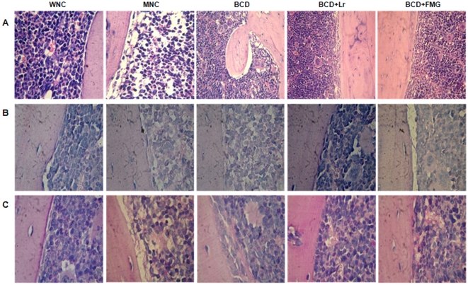
Figure 3. Histological examination of BM architecture from mice well-nourished control (WNC) mice, malnourished control (MNC) mice, and malnourished mice replete for 7 days with a balanced conventional diet (BCD) with supplemental Lactobacillus rhamnosus (BCD+Lr) or fermented goat milk (BCD+FGM).
The femur was removed; the BM fixed in paraformaldehyde, decalcified in formic acid and sodium citrate, and stained with (A) Hematoxylin and eosin, (B) Alcian Blue or (C) Periodic Acid Schiff. Light micrographs, original magnification ×400.

