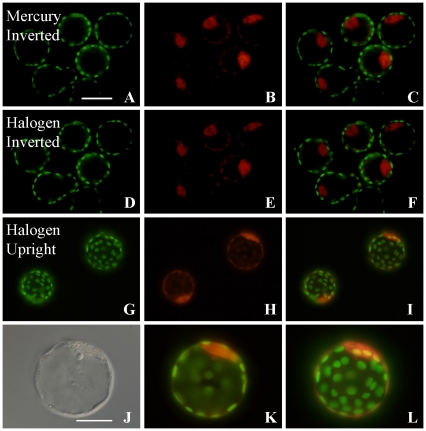
Figure 1. Observation of fluorescent dyes using a halogen lamp.
Fixed mouse blastocysts were treated with anti-Cdx2- and anti-Oct3/4-antibodies and stained with Alexa Fluor 488 (green) and 548 (red), respectively, then observed using a mercury vapor lamp (A–C) or an inverted microscope with a halogen lamp (D–F). Although the images produced by halogen light illumination were slightly weaker than images produced with the mercury vapor lamp, they could substitute for those seen using a traditional fluorescence microscope. Similar specimens were observed using an upright microscope with halogen lamp (G–L). These images were taken using an LCPlanF1 objective lens (×20; bar = 100 µm). (J) Bright field illumination. (K, L) A different focal plane of the same embryo shown in (I). (J) Inner cell mass (ICM) cells appear as Oct3/4-positive cells. (K, L) Merged images. (J–L) Observed using an LCPlanF1 objective lens (×40; bar = 50 µm).

